
Understanding Bone Grafting Procedures
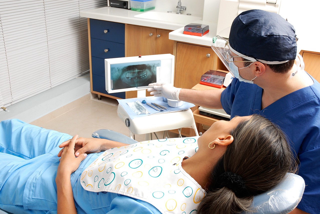
Implant dentistry has transformed the landscape of tooth replacement, offering patients durable and aesthetically pleasing solutions. However, successful implant placement often relies on adequate bone volume and density for osseointegration. In cases where the alveolar ridge lacks the necessary dimensions due to tooth loss, trauma, or resorption, the best dentist Redwood City may recommend bone grafting. Augmenting the deficient ridge with bone graft materials facilitates implant placement, ensuring stability and longevity.
Here are common reasons why individuals may require these procedures to restore oral health and function:
Insufficient Bone Volume for Implant Placement
One of the indications for bone grafting in dentistry is the inadequate volume or density of bone at the site of dental implant placement. Following tooth loss, bone resorption often occurs, leading to a reduction in bone height and width. Without adequate bone support, implant stability and long-term success are compromised. Therefore, bone grafting procedures are necessary to augment the deficient bone and create a suitable foundation for implant placement, ensuring optimal osseointegration and prosthetic stability.
Ridge Augmentation for Prosthetic Support
Patients seeking dental prostheses such as fixed or removable dentures, may require ridge augmentation procedures due to the resorption or deformities of the alveolar ridge. Insufficient ridge width or height can impair the retention and stability of prosthetic appliances, compromising oral function and aesthetics. By augmenting the ridge with bone graft materials, the best dentist can recreate a more anatomically favorable ridge morphology, enhancing the stability and longevity of dental prostheses and improving patient comfort and satisfaction.
Management of Periodontal Defects
Periodontal diseasescan result in the loss of periodontal tissues. Advanced periodontal defects, such as furcation defects, pose significant challenges in maintaining tooth stability and supporting structures. Bone grafting procedures are essential for regenerating lost periodontal tissues and promoting new bone formation. By restoring bone support around affected teeth, bone grafting aids in preserving tooth function and enhancing periodontal health, improving overall oral hygiene, and preventing further tooth loss.
Preservation of Extraction Sockets
Following tooth extraction, the alveolar bone undergoes resorption, leading to a reduction in bone volume and alterations in ridge morphology. Socket preservation techniques, involving the placement of bone graft materials into the extraction socket, help maintain the dimensions of the alveolar ridge and minimize bone loss. Preserving the extraction socket through bone grafting facilitates future implant placement and enhances the esthetic outcomes of dental treatments.
Biological Mechanisms of Bone Graft Integration
The success of bone grafting relies on the integration of graft materials with the host bone tissue. This integration performed by the best dentist is facilitated by complex biological mechanisms involving osteoconduction, osteoinduction, and osteogenesis.
Osteoconduction. It is a fundamental process in bone graft integration that involves the guiding of new bone formation along the surface of the graft material. The graft material serves as a scaffold or framework onto which osteogenic cellssuch as osteoblasts, can migrate and proliferate. Porous structures within the graft material allow for the infiltration of blood vessels and progenitor cells from the surrounding host tissue. As these cells populate the graft surface, they deposit new bone matrix, gradually bridging the gap between the graft and host bone.
Osteoinduction. It is a process by which certain graft materials possess the ability to stimulate the differentiation of undifferentiated mesenchymal stem cells into osteoblasts, thereby promoting bone formation. This phenomenon is mediated by the release of bioactive molecules, such as bone morphogenetic proteins (BMPs), growth factors, and cytokines, from the graft material.
These signaling molecules initiate a cascade of cellular events that culminate in the recruitment, proliferation, and differentiation of osteogenic cells within the graft site. Osteoinductive graft materialsand certain bone morphogenetic protein (BMP)-based products, help in bone regeneration and graft integration.
Osteogenesis. This refers to the formation of new bone tissue by osteogenic cells derived from either the host tissue or the graft material itself. In autogenous bone grafting, the graft material contains viable osteogenic cells, which contribute to bone formation directly. These cells undergo proliferation, differentiation, and mineralization processes, leading to the formation of new bone tissue.
In contrast, allografts and xenografts lack viable osteogenic cells and rely on osteoconduction and osteoinduction mechanisms to facilitate bone regeneration. However, they still provide an osteoconductive scaffold for host-derived osteogenic cells to populate and form new bone.
Innovations in Bone Grafting Technology
Advancements in bone grafting technology have transformed the landscape of bone regeneration and reconstruction. These innovative approaches offer the best dentist more effective tools for promoting bone healing, enhancing graft integration, and improving bone grafting procedures.
Growth Factors.Growth factors regulate cellular activities involved in bone healing and regeneration. Recent advancements in biotechnology have enabled the development of recombinant growth factors that mimic the natural signaling pathways involved in bone formation. These growth factors are often incorporated into bone graft materials or delivered locally to the graft site, enhancing bone healing and accelerating the integration of graft materials with the host bone tissue.
Stem Cell Therapy. This therapy holds immense promise in the field of bone regeneration, offering a regenerative approach to treating bone defects and deficiencies. Mesenchymal stem cells (MSCs) derived from various sources such as bone marrow, adipose tissue, and dental pulp, have the ability to differentiate into osteoblasts and contribute to bone formation.
In bone grafting procedures, MSCs can be isolated, expanded in vitro, and combined with scaffold materials to create tissue-engineered constructs. The best dentistimplants these constructs into the defect site, where MSCs facilitate bone regeneration by secreting growth factors, modulating the immune response, and promoting angiogenesis.
3D-Printed Scaffolds.3D printing technology has revolutionized the fabrication of customized scaffolds for bone tissue engineering. By controlling the design parameters such as pore size, porosity, and mechanical properties, 3D-printed scaffolds can be tailored to match the anatomical features of the defect site and provide optimal support for bone regeneration. These scaffolds are typically composed of biocompatible materials, which promote cell adhesion, proliferation, and tissue ingrowth.
In addition, bioactive agentssuch as growth factors or antimicrobial agents, can be incorporated into the scaffold matrix to further improve bone healing. 3D-printed scaffolds offer the best dentist a versatile and customizable solution for addressing complex bone defects and achieving precise surgical outcomes.